EYE (cont'd)
Rods vs. Cones
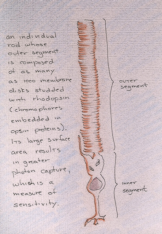
All rods are primed with the same protein-opsin complex and are tuned therefore to the same wavelength of light. This uniform sensitivity, however, is an obstacle to their employ in the neuronal activities required for the psychological confection of color. That said, rods have long outer segments containing a thousand or so discs whose resulting surface area can accomodate hundreds of thousands of pigment molecules. So, the probability of photon catch is greater--significantly greater--for rods than for cones, rendering them peak-performers in scotopic or dark circumstance.
But low light--say, light streaming from a star many millions of lightyears away--will impart only a little energy to the rod, its photons striking only a small number of chromophores at an understandably infrequent rate. The limited photon catch of this rod is insufficient to cause the ganglion cell at the end of the chain to pulse. But that is not necessarily a barrier to your viewing of the star. Rods commonly pool their signals such that weak signals can then be combined into a single strong signal as the various rods within a given receptive field all converge upon an individual bipolar cell. The bipolar cell is then able to pass on this summated energy/signal to the ganglion cell, which now can pulse and signal the brian.
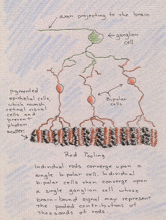
In view of the preceding, we will generalize by stating that the rod has come to assume a design that favors sensitivity over acuity. By pooling signals from a given receptive field, rods enable the retina to register otherwise forbiddingly weak or remote signals. And then, again, by pooling signals from a given receptive field, rods confuse the possibility of determining from which particular point within that given receptive field the signal emanates. For, in fact, it emanates from many points and is only a single signal by virtue of pooling or compression.
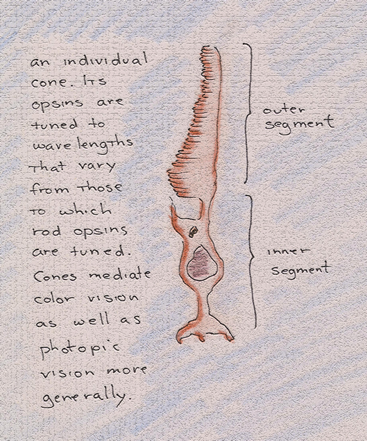
Cones, on the other hand, are of a design and function to favor acuity over sensitivity. They are found, for the most part, in and around the fovea. Their conically-shaped outer segment is shorter than that of the rod and, as a result, they have less surface area and are thus responsible for a much smaller photon catch.
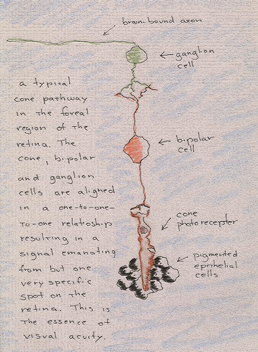
Their sensitivity is further compromised by the fact that cones do not pool or compress their signals as do rods. In the foveola for example, the very center of the fovea, cones synapse with bipolar cells, and bipolar cells with ganglion cells, in a one-to-one relationship. So, while cones require photopic or lighted circumstance in which to function, they nevertheless exhibit fidelity to the point from which the signal emanantes on the retina. They are, in other words, the mediators of visual acuity or of that fineness of vision associated with the center of view.
Cones are also distinguished from rods for the fact that not all cones have the same protein-opsin complex embedded within the membrane invaginations of their outer segments. Some cones have protein variants that tune their opsins to short-wave light; some, to medium-wave light; and some, to long-wave light.
The varying sensitivities of S-cones, M-cones and L-cones are instrumental in our perception of color but--and this is an important caveat--they do not signal the brain as to which wavelength they are responding to. Their signals are no different from the signals sent to the brain by rods: the ganglion cell representative will pulse to indicate that the receptor has been exposed to light and that pulse will vary only in frequency--not height or length--in accordance with the duration or intensity of the exposure.
[Our apologies as this section is still to be edited]
Acuity vs. Sensitivity
Dark Current
Color (Spectral Sensitivities)
